
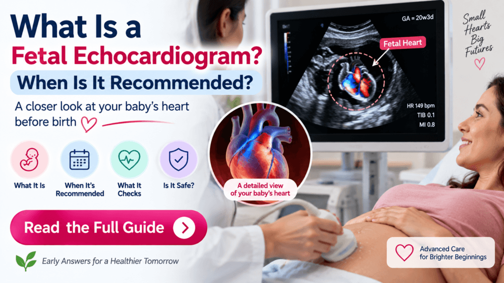
A fetal echocardiogram, sometimes called a fetal echo, is a specialized ultrasound examination that looks closely at a developing baby’s heart before birth. Unlike the basic heart screening included in a routine pregnancy ultrasound, a fetal echocardiogram provides a more detailed assessment of the heart’s structure, rhythm, blood flow, and function.
For most pregnancies, a routine anatomy ultrasound is enough to screen the baby’s heart. A fetal echocardiogram is usually recommended when there is a higher-than-usual risk of congenital heart disease or when the routine ultrasound shows a possible heart abnormality.
A fetal echocardiogram is a detailed prenatal ultrasound of the baby’s heart. It is most commonly performed around 18–22 weeks of pregnancy.
Doctors may recommend it if a routine ultrasound shows a possible heart problem, there is a family history of congenital heart disease, the mother has certain medical conditions such as pregestational diabetes, or the fetus has other findings associated with an increased risk of heart disease
What Is a Fetal Echocardiogram?
A fetal echocardiogram is a specialized ultrasound examination of the fetal heart.
It uses sound waves to create detailed, moving images of the baby’s heart while the baby is still in the uterus. The examination can evaluate the heart’s chambers, valves, major blood vessels, rhythm, and aspects of cardiac function.
It is considered the specialized diagnostic test used to evaluate suspected congenital heart disease before birth. The 2023 American Society of Echocardiography (ASE) guideline describes fetal echocardiography as the gold-standard test for diagnosing congenital heart disease in the fetus.
How Is a Fetal Echo Different From a Regular Ultrasound?
A standard anatomy ultrasound includes screening views of the fetal heart. Depending on the practice and image quality, these may include the four-chamber view and views of the heart’s outflow tracts and major vessels.
A fetal echocardiogram goes much further.
During a dedicated fetal echo, the specialist can examine:
- Four heart chambers
- Heart valves
- Outflow tracts
- Aortic and ductal arches
- Major blood vessels
- Venous connections
- Heart rhythm
- Blood flow
- Cardiac function
The examination uses multiple ultrasound techniques, including 2D imaging, M-mode, color Doppler, and pulsed-wave Doppler when appropriate.
When Is a Fetal Echocardiogram Recommended?
The most important reason for a fetal echocardiogram is an abnormal or concerning finding on the routine fetal heart screening ultrasound.
Also Read : Echogenic Intracardiac Focus (EIF) on Ultrasound: What Does It Mean?
ASE guidance states that fetal echocardiography is indicated when the cardiac screening ultrasound is abnormal, regardless of whether other risk factors are present.
A fetal echo may also be recommended when certain maternal, fetal, or family factors increase the likelihood of congenital heart disease.
Common Reasons Include:
- A suspected heart abnormality on routine ultrasound
- A parent or sibling with congenital heart disease
- Pregestational diabetes
- Maternal phenylketonuria (PKU)
- Certain maternal autoimmune conditions, particularly with SSA/SSB antibodies
- Certain maternal infections, such as rubella
- Certain medication exposures
- Abnormal fetal heart rhythm
- Fetal hydrops
- Another fetal abnormality
- Certain chromosomal abnormalities
- Monochorionic twins
- Increased nuchal translucency
- Some pregnancies conceived using IVF or assisted reproductive technology
The exact indications can differ depending on the patient’s individual risk and local clinical practice.
Who May Need a Fetal Echo?
Family History of Congenital Heart Disease
If a parent or sibling has congenital heart disease, the baby’s risk may be higher than the general population. A fetal echocardiogram is generally recommended when there is a first-degree family history of congenital heart disease.
Pregestational Diabetes
Diabetes that existed before pregnancy is an established indication for fetal echocardiography in current ASE recommendations.
This is different from gestational diabetes diagnosed later in pregnancy. ASE specifically distinguishes pregestational diabetes from gestational diabetes diagnosed after the second trimester when considering fetal echo referral.
Abnormal Routine Ultrasound
If the anatomy scan shows an abnormality involving the fetal heart—such as an unusual chamber appearance, abnormal outflow tract, or other concerning finding—a dedicated fetal echocardiogram may be recommended.
This is one of the strongest indications for referral.
Abnormal Fetal Heart Rhythm
If the baby’s heart rate is unusually fast, slow, or irregular, a fetal echocardiogram may be used to evaluate the rhythm and look for associated cardiac abnormalities.
Increased Nuchal Translucency
An increased nuchal translucency measurement can be associated with an increased risk of congenital heart disease. ASE recommendations consider fetal echocardiography for nuchal translucency measurements of 3.5 mm or greater and say it may be considered for measurements of 3.0–3.4 mm, depending on the clinical context.
IVF or Assisted Reproductive Technology
The ASE 2023 guideline says fetal echocardiography may be considered for pregnancies conceived through IVF, while other organizations may have somewhat different recommendations. The decision can depend on the quality of the routine cardiac screening and other risk factors.
When Is the Best Time to Have a Fetal Echocardiogram?
The optimal timing for a comprehensive transabdominal fetal echocardiogram is generally 18–22 weeks of pregnancy. This overlaps with the timing of the detailed anatomy ultrasound.
In selected high-risk pregnancies, fetal cardiac imaging may be performed earlier, sometimes around 12–14 weeks. However, early examinations have limitations, and ASE recommends that early studies generally be repeated later in the second trimester.
Also Read : Placenta Is Low-Lying at 20 Weeks: What Happens Next?
Therefore, having an early fetal echo does not necessarily mean that a second examination will be unnecessary.
What Happens During the Test?
Most fetal echocardiograms are performed through the mother’s abdomen.
A sonographer places ultrasound gel on the abdomen and moves an ultrasound transducer over the skin. Detailed images of the fetal heart are obtained from multiple angles.
The test is:
- Noninvasive
- Generally painless
- Performed without needles
- Similar to an ultrasound examination
- Usually performed while the mother is awake
The American Heart Association reports that a fetal echocardiogram can take approximately 30 minutes to 2 hours, depending on how detailed the examination needs to be and how easy it is to obtain the required images.
The images may then be reviewed by a specialist with expertise in fetal and congenital heart disease.
What Does a Fetal Echocardiogram Check?
A fetal echo provides a detailed assessment of the baby’s cardiovascular system.
The examination may evaluate:
Heart Chambers
The specialist examines the four chambers of the heart and their size, shape, and relationships.
Heart Valves
The valves between the chambers are evaluated for their appearance and movement.
Outflow Tracts
The pathways through which blood leaves the heart are examined, including the major arteries.
Aortic and Ductal Arches
These major vessels are assessed for their anatomy and blood flow.
Heart Rhythm
The fetal heart rate and rhythm can be evaluated for abnormalities such as certain arrhythmias.
Blood Flow
Doppler ultrasound can provide information about blood flow through different parts of the fetal heart and circulation.
Cardiac Function
The specialist can assess how the heart is functioning and look for signs of cardiovascular stress or dysfunction.
Is a Fetal Echocardiogram Safe?
Fetal echocardiography uses ultrasound rather than X-rays or radiation.
When performed for an appropriate medical indication using recommended ultrasound safety practices, it is considered a safe prenatal imaging examination. The AIUM/ACOG/SMFM-endorsed practice guidance emphasizes using the lowest ultrasound exposure settings necessary to obtain the required diagnostic information.
You generally do not need special preparation for a fetal echocardiogram, although instructions can vary by facility. The American Heart Association notes that a full bladder is not usually required.
What Can a Fetal Echo Detect?
A fetal echocardiogram can detect or help characterize many significant congenital heart abnormalities before birth.
Examples include:
- Abnormal heart chamber development
- Abnormal connections between chambers or vessels
- Certain valve abnormalities
- Outflow tract abnormalities
- Some forms of congenital heart disease
- Abnormal fetal heart rhythms
- Certain problems affecting cardiac function
Prenatal diagnosis can be particularly valuable when a serious heart defect could affect delivery location, newborn stabilization, medication, or surgery after birth.
However, a normal fetal echocardiogram cannot rule out every possible heart problem. Some small defects or abnormalities that develop later may be difficult to identify before birth. The American Heart Association specifically notes that fetal circulation differs from circulation after birth and that some small heart problems can be difficult to detect prenatally.
What Happens if the Result Is Abnormal?
An abnormal fetal echocardiogram does not necessarily mean the baby will have a severe medical problem.
The next step depends on the specific finding.
You may be referred to a pediatric cardiologist, maternal-fetal medicine specialist, genetic counselor, or other specialist for additional evaluation and counseling.
Depending on the diagnosis, doctors may recommend:
- Repeat fetal echocardiograms
- Additional ultrasound examinations
- Genetic counseling or testing
- Additional monitoring during pregnancy
- Delivery at a specialized medical center
- A newborn cardiology evaluation
- Planning for treatment after birth
Some fetal heart conditions require serial monitoring because they can change as pregnancy progresses. ASE guidance notes that follow-up intervals depend on the specific heart lesion and clinical concerns.
When to Contact Your Doctor
A fetal echocardiogram itself is not usually an emergency test. However, contact your pregnancy care provider if:
- Your routine ultrasound showed a possible heart abnormality.
- Your baby’s heart rate was unusually fast, slow, or irregular.
- Your doctor recommended a fetal echocardiogram but you are unsure why.
- You have a significant family history of congenital heart disease.
- You have a medical condition associated with increased fetal cardiac risk.
- You have questions about an abnormal fetal echo result.
If you develop urgent pregnancy symptoms such as heavy bleeding, severe abdominal pain, fluid leakage, fainting, or other emergency symptoms, seek prompt medical attention.
Is a fetal echocardiogram the same as a regular ultrasound?
No. A fetal echocardiogram is a specialized, detailed examination focused specifically on the baby’s heart, while a routine anatomy ultrasound evaluates the entire fetus and includes basic cardiac screening.
When is a fetal echocardiogram usually done?
The standard window for a comprehensive fetal echocardiogram is 18–22 weeks of pregnancy. Earlier studies may be performed in selected high-risk situations.
Why would my doctor recommend a fetal echo?
Common reasons include an abnormal heart finding on ultrasound, a family history of congenital heart disease, pregestational diabetes, certain maternal conditions or exposures, abnormal fetal heart rhythm, increased nuchal translucency, or certain fetal abnormalities.
Does IVF mean I definitely need a fetal echocardiogram?
Not necessarily. IVF is considered an indication that may warrant fetal echocardiography under the 2023 ASE recommendations, but referral decisions can depend on other risk factors and the quality of the routine cardiac screening.
What if the fetal echocardiogram is normal?
A normal fetal echo is reassuring and usually means no significant cardiac abnormality was identified during the examination. However, no prenatal test can detect every possible heart problem.
Can a fetal heart problem be treated before birth?
In selected situations, fetal cardiac conditions can be managed or treated during pregnancy. However, most congenital heart defects are managed through careful monitoring and planning for delivery and treatment after birth.

