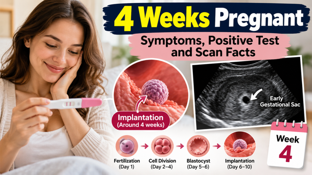

At 3 weeks pregnant, the most important events of early life take place, yet there is almost nothing to see or feel. An egg is fertilized, the new cell begins dividing, and a tiny ball of cells starts its journey toward your uterus, where it will attach in a process called implantation. If you are wondering what an ultrasound would show at this stage, or what a 3 week ultrasound report might say, the short answer is that a scan cannot see fertilization or implantation, and a normal report at this point usually reads “no gestational sac seen”. This guide explains fertilization and implantation step by step, what an ultrasound can and cannot show at 3 weeks, how to read the common phrases in an early ultrasound report, and when the first real signs appear on a scan.
Series navigation: Previous week: 1 Week Pregnant and 2 Weeks Pregnant | Next week: 4 Weeks Pregnant
What Does 3 Weeks Pregnant Mean?
Doctors count pregnancy from the first day of your last menstrual period, not from the day of conception. In weeks 1 and 2 you are not pregnant yet. Your period ends, your uterine lining rebuilds, and an egg matures in an ovary. Conception usually happens between weeks 2 and 3, so week 3 is the first week in which a pregnancy truly exists. Because the embryo is only days old, the age of the baby is about two weeks less than the number of weeks you are counted as pregnant.
Important: At 3 weeks pregnant, a home pregnancy test and an ultrasound will usually both be negative or non-diagnostic. This does not mean anything is wrong. It simply means the pregnancy is too early to detect.
Fertilization Explained
Fertilization begins when ovulation releases an egg, which is picked up by the fallopian tube. The egg lives for only about 12 to 24 hours, so fertilization must happen quickly. Sperm can survive in the reproductive tract for up to five days, which is why sex in the days before ovulation can lead to pregnancy. When a sperm penetrates the egg, the two sets of DNA combine into one cell called a zygote, and the baby’s genetic blueprint is set. This includes traits such as eye color, hair color, and sex.
The zygote then begins dividing, first into two cells, then four, then eight, and so on, while it moves slowly down the fallopian tube. After several days it becomes a solid ball of cells, and later a hollow ball called a blastocyst. All of this happens at a microscopic level, with a structure far smaller than the point of a pin.
Implantation Explained
Implantation is the moment the blastocyst attaches to the lining of the uterus, which has been thickened by progesterone to receive it. It usually happens about 6 to 10 days after fertilization, which is around the end of week 3 or during week 4 of pregnancy. The outer cells of the blastocyst burrow into the lining and begin forming connections to your blood supply. These cells also start producing human chorionic gonadotropin (hCG), the hormone that home pregnancy tests detect. After implantation, a yolk sac develops and nourishes the embryo until the placenta takes over.
Some people notice light spotting or mild cramping around implantation, often called implantation bleeding. It is usually pink or brown, light, and short, lasting from a few hours to about two days. Many people notice nothing at all, and neither experience is a sign of a problem.
| Stage | Approximate timing | Can an ultrasound see it? |
|---|---|---|
| Ovulation | About day 14 of a 28-day cycle | The dominant follicle can be seen before it ruptures |
| Fertilization | Within about 24 hours of ovulation | No, it is microscopic |
| Cell division and blastocyst | First 5 days after fertilization | No |
| Implantation | About 6 to 10 days after fertilization | No, but the lining may look thick |
| Gestational sac | Around 4.5 to 5 weeks of pregnancy | Yes, on a transvaginal scan |
What Does an Ultrasound Show at 3 Weeks Pregnant?
At 3 weeks pregnant, an ultrasound cannot show the embryo, the fertilized egg, or implantation, because these structures are far too small to be resolved. The first structure that can be seen in the uterus is the gestational sac, and it appears at about 4.5 to 5 weeks of pregnancy on a transvaginal scan. If a scan is done at 3 weeks, it can only show the organs and the indirect changes that surround early pregnancy.
Also Read : How Early Can an Ultrasound Detect Pregnancy? Everything You Need to Know
Typical findings include a normal-sized uterus with a thickened, bright endometrium, which is the lining prepared by progesterone after ovulation. One ovary often shows a small cyst-like structure called a corpus luteum, which forms from the ruptured follicle and produces progesterone to support the early pregnancy. There is usually no free fluid, and no mass in the tubes or ovaries. None of these findings can confirm a pregnancy by themselves, because the same appearance is seen in the second half of any normal menstrual cycle.
What the Ultrasound Report May Say at 3 Weeks
If you do have a scan at 3 weeks, usually because of pain, spotting, or concerns about dates, the report will use standard phrases. The table below explains the most common ones in plain language.
| Phrase in the report | What it means |
|---|---|
| Uterus anteverted, normal size and shape | The uterus tilts forward, which is the most common position, and looks normal |
| Endometrial thickness (for example 10 to 15 mm), secretory or thickened | The lining is thick and prepared, as it is after ovulation, whether or not you are pregnant |
| No intrauterine gestational sac seen | No sac is visible yet, which is expected at this early stage |
| Corpus luteum seen in the ovary | The follicle that released the egg has formed a normal hormone-producing structure |
| No adnexal mass, no free fluid | The tubes and ovaries look normal and there is no fluid in the pelvis |
| Pregnancy of unknown location (PUL) | The hCG test is positive but the scan shows no pregnancy yet, so follow-up is needed |
| Advised: serum beta-hCG and repeat scan in 7 to 10 days | The usual next step to confirm where the pregnancy is and how it is developing |
The wording and numbers differ from lab to lab, so always ask your doctor to explain your own report.
Important: “No gestational sac seen” at 3 weeks is normal and does not mean a miscarriage. If you also have a positive hCG test with pain or bleeding and no sac is seen, your doctor will arrange follow-up quickly, because an ectopic pregnancy must be ruled out.
When Can Ultrasound First See the Pregnancy?
The gestational sac is the first structure seen, at 4.5 to 5 weeks on a transvaginal scan, and a transabdominal scan usually sees it closer to 6 weeks. The yolk sac, a small bright ring inside the sac, appears at about 5.5 weeks. The embryo itself, a 1 to 2 millimeter structure beside the yolk sac, is seen at about 6 weeks, and the heartbeat appears as a flicker around the same time. A hormone level called beta-hCG helps interpret the scan. When it passes a threshold usually described as 1,500 to 2,000 mIU/mL, and as high as 3,500 in some guidance, a normal intrauterine pregnancy is expected to be visible on a transvaginal scan.
Does a Routine Ultrasound Happen at 3 Weeks?
No. Without symptoms, there is no reason for a scan this early. In a low-risk pregnancy, the first trimester ultrasound is usually done between 11 and 13 weeks to confirm dating and check viability, and many providers also offer an earlier dating scan around 6 to 8 weeks. An early scan is more likely if you have pain, bleeding, a history of ectopic pregnancy, fertility treatment such as IVF, or uncertain dates. Ultrasound uses sound waves, not radiation, and is considered safe in pregnancy when performed by a trained professional.
Symptoms at 3 Weeks Pregnant
Most people feel nothing at 3 weeks pregnant. Mild cramps or light spotting may occur around implantation, but nausea, sore breasts, and tiredness at this stage are usually normal hormone shifts before a period and cannot be used to tell whether you are pregnant. A pregnancy test is the only reliable first step, and the best time to take one is on or after the day your period is due, using first morning urine.
What to Do at 3 Weeks Pregnant
Begin or continue a prenatal vitamin with at least 400 micrograms of folic acid, which the CDC recommends to help prevent neural tube defects. Avoid alcohol, tobacco, and recreational drugs, keep caffeine under 200 milligrams a day, check all medicines with your provider, eat a balanced diet, and follow food safety basics. Once you have a positive test, call your provider to schedule your first prenatal visit.
When to Call Your Doctor
Call your provider right away if you have heavy bleeding, severe or one-sided pelvic pain, shoulder-tip pain, dizziness, or fainting. These can signal an ectopic pregnancy, where the embryo implants outside the uterus, which needs urgent care.
Final Thoughts on 3 Weeks Pregnant
At 3 weeks pregnant, fertilization has happened and implantation is about to begin, but both are invisible to ultrasound. A scan at this stage mostly shows a thickened lining and a corpus luteum, and a report that says no gestational sac seen is normal for the timing. The first real ultrasound evidence of pregnancy arrives at 4.5 to 5 weeks, followed by the yolk sac and the heartbeat. Focus on folic acid and healthy habits now, take a test when your period is due, and let your provider decide whether and when a scan is needed.
This article is for educational and informational purposes only and is not a substitute for professional medical advice, diagnosis, or treatment. Always consult a qualified healthcare professional for advice about your individual health.