
At 7 weeks pregnant, your baby is about the size of a blueberry, roughly 1 centimeter from head to bottom, and growing at a remarkable pace. The tiny heart is beating about twice as fast as yours, arm and leg buds are lengthening, and a face is beginning to take shape. Meanwhile, your body is working hard, and symptoms such as nausea, tiredness, and sore breasts are often at their strongest. If you have an ultrasound this week, you will probably see a C-shaped embryo with a flickering heartbeat, and you may leave with a report full of unfamiliar terms. This guide explains your baby’s size and development at 7 weeks pregnant, the symptoms to expect, what a 7-week ultrasound shows, how to read the report, and when to call your doctor.
What Is Happening at 7 Weeks Pregnant?
Pregnancy is counted from the first day of your last menstrual period, so at 7 weeks pregnant the embryo is about five weeks old. This is the middle of the second month of pregnancy and a time of fast change. The placenta is developing and gradually taking over the job of feeding your baby, which until now has been done largely by the yolk sac. Your hormone levels remain high, your blood volume is beginning to rise, and your uterus is growing to about the size of a lemon, even though you still look the same from the outside.
Important: If your cycles are irregular or you ovulated late, your baby may be a few days behind your calendar dates, which is why doctors use the embryo’s measured size to confirm your due date.
Baby Size at 7 Weeks Pregnant
At 7 weeks, a baby is usually compared to a blueberry and measures about 1 centimeter (roughly half an inch) from the top of the head to the bottom, which is called the crown-rump length (CRL). Doctors use the CRL, instead of the full length, because the embryo is curled up in a C shape. Published figures vary slightly, and a typical range for 7 weeks is about 6 to 15 millimeters, because the embryo grows about a millimeter a day and a few days make a visible difference. In the first trimester, the CRL is the most accurate way to date a pregnancy, with an accuracy of about five days.
Baby Development at 7 Weeks Pregnant
| Body part | What is happening at 7 weeks |
|---|---|
| Heart | The heart keeps developing and beating at roughly 100 to 160 beats per minute |
| Brain | The brain grows quickly and divides into more defined regions |
| Face | Nostrils, the lens of the eyes, and the mouth are beginning to form |
| Arms and hands | The arm buds are lengthening and flattening into paddle-shaped hands |
| Legs and feet | The leg buds are following, slightly behind the arms |
| Organs | The liver, kidneys, lungs, and digestive tract continue to develop |
| Umbilical cord | The cord that connects your baby to the placenta is forming |
At this stage the embryo may still show a small tail-like structure, which is normal and disappears as development continues. Folic acid remains important because the brain and spinal cord are still developing, so keep taking a prenatal vitamin, and remember that the CDC recommends at least 400 micrograms of folic acid daily.
Your Baby’s Heartbeat at 7 Weeks
By 7 weeks, the heart has been beating for about a week or two, and it is easier to see on an ultrasound than it was at 6 weeks. The heart rate rises quickly in early pregnancy. One study of 255 pregnancies found an average embryonic heart rate of about 116 beats per minute at 6 weeks, increasing to about 172 at 10 weeks. At 7 weeks, many sources describe a normal range of roughly 100 to 160 beats per minute, and numbers vary a little between studies and ultrasound machines. On screen, the heartbeat appears as a steady flicker, since the embryo is too small for a stethoscope to detect. A single reading is not a verdict, because the rate changes every day and depends on exactly how many days along you are.
What Does a 7-Week Ultrasound Show?
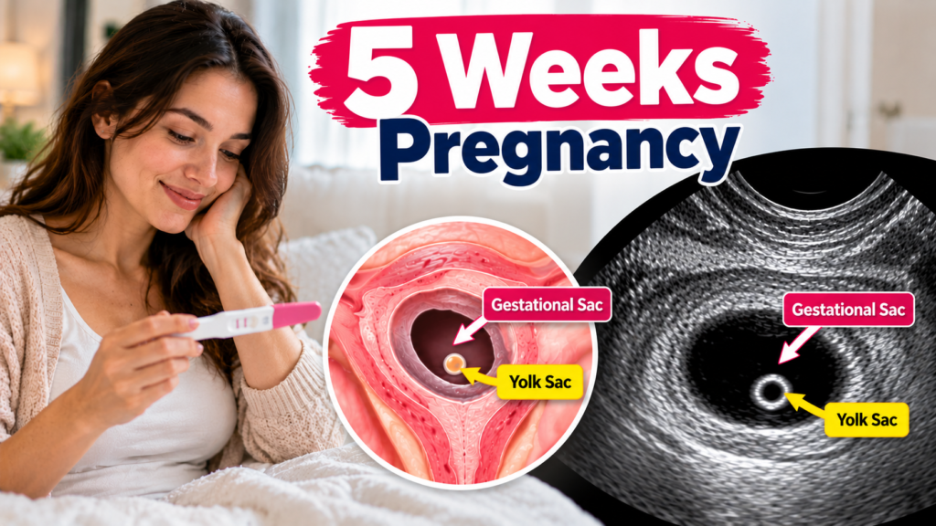
A scan at 7 weeks is almost always done transvaginally, because the probe sits close to the uterus and gives a clearer picture than an abdominal scan can at this size. The sonographer checks several things. First, they confirm the pregnancy is inside the uterus, which helps rule out an ectopic pregnancy. Then they look at the gestational sac, the fluid-filled space the embryo grows in, and the yolk sac, a small bright ring next to the embryo. The embryo itself appears as a thick, curved shape, and the heartbeat is checked and the rate recorded. The sonographer measures the CRL to confirm how far along you are, and counts the number of embryos, because twins can be identified at this stage. At about 7 weeks, a very thin membrane called the amnion often becomes visible around the embryo.
The image will not look like a baby. At 7 weeks, you will see a small, C-shaped bean with a flickering light, and that is exactly what a healthy 7-week pregnancy looks like. Early scans use sound waves, not radiation, and are considered safe in pregnancy when performed by trained professionals. A routine scan is not always offered this early, but one is commonly done if you have pain or bleeding, are having fertility treatment, have a history of ectopic pregnancy, or are unsure of your dates.
6 Weeks 3 Days Ultrasound: What You Will Actually See on the Screen
How to Read Your 7-Week Ultrasound Report
| Phrase in the report | What it means |
|---|---|
| Single live intrauterine pregnancy | One embryo with a heartbeat is growing inside the uterus |
| Gestational sac, mean sac diameter (MSD) in mm | The size of the sac, which should grow steadily |
| Yolk sac seen | The yolk sac is present, which is a reassuring early sign |
| Crown-rump length (CRL) in mm, corresponding to 7 weeks and a few days | The embryo’s measured size and the age it suggests |
| Cardiac activity present, FHR in bpm | A heartbeat was seen, and the heart rate was recorded |
| EDD by CRL | The estimated due date based on the embryo’s size |
| Subchorionic hematoma or hemorrhage | A small collection of blood near the sac that is fairly common and often resolves |
| Corpus luteum cyst | A normal hormone-producing cyst on the ovary |
| No adnexal mass, no free fluid | The tubes and ovaries look normal |
The exact wording differs between clinics, so ask your doctor to explain your report in the context of your symptoms and tests.
Tip: If your CRL gives a slightly different date from your last period, your doctor may adjust your due date. This is routine and does not mean anything is wrong.
What If the Scan Is Behind or No Heartbeat Is Seen?
Most scans at 7 weeks show a heartbeat, but not all, and a result that is less clear than hoped is not automatically bad news. Dating can be off by a week, and a smaller embryo may simply be younger than expected. Guidelines are careful for this reason. A heartbeat is expected once the CRL reaches about 7 millimeters. When the embryo is smaller than that and no heartbeat is seen, doctors typically repeat the scan at least a week later. If the embryo is 7 millimeters or more with no heartbeat, doctors usually confirm the finding with a second opinion or repeat scan before giving a diagnosis. If your results are uncertain, your care team will explain the next steps, and it is reasonable to ask questions at every step.
Important: Call your doctor right away if you have heavy bleeding, severe or one-sided abdominal pain, shoulder pain, dizziness, or fainting, because these can signal an ectopic pregnancy or another emergency.
Symptoms at 7 Weeks Pregnant
| Symptom | What to know |
|---|---|
| Nausea and vomiting | Often strong now, and it can happen at any time of day |
| Tiredness | Rising progesterone and blood volume can leave you exhausted |
| Breast changes | Breasts may feel heavy, tender, and swollen |
| Frequent urination | The growing uterus presses on the bladder |
| Food aversions and cravings | Strong smells and tastes can set off nausea |
| Heartburn, bloating, constipation | Hormones slow digestion |
| Excess saliva or sore gums | Some people notice more saliva or bleeding gums |
| Mood swings | Common as hormone levels change |
To ease nausea, eat small, frequent meals, keep plain crackers nearby, sip fluids through the day, and avoid triggers. Some people find ginger helpful, and ACOG recommends vitamin B6, with or without doxylamine, as the first-line medicine, so ask your provider before using anything. Most people gain little weight in the first trimester, often about 1 to 5 pounds, and some lose a little because of nausea. Call your provider if you cannot keep fluids down, are losing weight, or feel dizzy, because severe vomiting may need treatment.
What to Do at 7 Weeks Pregnant
Book your first prenatal visit if you have not done so, since many providers see patients around 8 weeks, and expect blood tests, a review of your history, and possibly an ultrasound. Continue a daily prenatal vitamin with folic acid, avoid alcohol, tobacco, and drugs, and keep caffeine under 200 milligrams a day. Check every medicine and supplement with your provider, practice food safety by cooking meat and eggs well and avoiding unpasteurized dairy, and stay hydrated. Gentle walking is usually fine if your provider agrees. Rest when you can, and consider telling someone you trust how you feel, since early pregnancy can be exciting and stressful at the same time.
Placenta Maturity Grades 0,1,2 & 3 Explained: How Each Stage Affects Your Baby
Final Thoughts on 7 Weeks Pregnant
At 7 weeks pregnant, your baby is the size of a blueberry, with a beating heart, growing limb buds, and the beginnings of a face. A 7-week ultrasound usually shows the gestational sac, yolk sac, a C-shaped embryo, and a heartbeat in the range of roughly 100 to 160 beats per minute, and the CRL confirms your due date. If the picture is less clear than you hoped, a repeat scan in about a week is the usual next step. Take your prenatal vitamin, rest, and keep your appointments. Next week, you will learn what changes at 8 weeks pregnant.
This article is for educational and informational purposes only and is not a substitute for professional medical advice, diagnosis, or treatment. Always consult a qualified healthcare professional for advice about your individual health.