
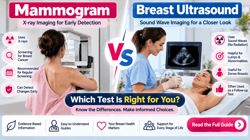
A mammogram and a breast ultrasound are both important breast-imaging tests, but they work differently and are used for different purposes. A mammogram uses low-dose X-rays, while a breast ultrasound uses sound waves to create images of breast tissue.
For routine breast cancer screening, mammography remains the primary imaging test. Breast ultrasound is commonly used to evaluate a breast lump or other abnormality and may also be used as supplemental imaging in selected women, including some women with dense breasts.
Quick Answer: Mammogram vs Breast Ultrasound
| Feature | Mammogram | Breast Ultrasound |
|---|---|---|
| Technology | Low-dose X-rays | Sound waves |
| Radiation | Yes, low-dose | No ionizing radiation |
| Main use | Breast cancer screening and diagnosis | Evaluating lumps and abnormalities |
| Dense breasts | May be less sensitive | Can show some findings hidden on mammography |
| Cysts vs solid masses | Sometimes less definitive | Often useful for distinguishing cystic vs solid lesions |
| Pregnancy | Requires special consideration | No ionizing radiation |
| Replaces mammography? | — | No |
| Can guide biopsy? | Yes, depending on the procedure | Yes |
Neither test is universally “better.” The appropriate examination depends on age, symptoms, breast density, pregnancy status, cancer risk, and the clinical question.
What Is a Mammogram?
A mammogram is a specialized breast-imaging examination that uses low-dose X-rays to create detailed images of the breast.
Mammography plays a central role in breast cancer screening because it can detect certain breast abnormalities before they can be felt or cause symptoms. It can also detect some forms of ductal carcinoma in situ (DCIS), including abnormalities that may appear as calcifications.
During a mammogram, the breast is positioned and gently compressed between plates while X-ray images are taken.
What Can a Mammogram Detect?
A mammogram may show:
- Masses
- Calcifications
- Architectural distortion
- Asymmetries
- Other suspicious breast changes
Mammography can be used for both screening and diagnostic evaluation.
What Is a Breast Ultrasound?
A breast ultrasound uses high-frequency sound waves to produce real-time images of breast tissue. It does not use ionizing radiation.
Also Read : What Is PCOS? Symptoms, Causes, Diagnosis & Treatment
During the examination, a sonographer or radiologist applies ultrasound gel to the breast and moves a transducer across the skin.
Breast ultrasound is particularly useful for evaluating a specific area of concern.
For example, ultrasound can help determine whether a breast lump is:
- Fluid-filled, such as a cyst
- Solid
- Complex or mixed cystic and solid
It can also characterize abnormalities found during a mammogram or physical examination.
Mammogram vs Ultrasound: Key Differences
1. Imaging Technology
The biggest difference is how the images are produced.
Mammogram: Uses low-dose X-rays.
Breast ultrasound: Uses sound waves.
Because ultrasound does not use ionizing radiation, it is particularly useful when radiation should be avoided or minimized, such as during pregnancy when imaging is clinically indicated.
2. Main Purpose
Mammography is primarily used for breast cancer screening and can also be used diagnostically.
Ultrasound is frequently used to investigate a lump, focal pain, nipple discharge, or an abnormality detected on another examination.
3. Breast Density
Dense breast tissue can make mammograms harder to interpret because both dense tissue and some cancers appear white on mammographic images.
Ultrasound can sometimes identify abnormalities that are difficult to see on mammography.
However, ultrasound has a higher false-positive rate and is not a replacement for mammography.
When Is a Mammogram Used?
Mammography is commonly used for routine breast cancer screening in women who do not have breast symptoms.
The exact screening schedule depends on the guideline and individual risk factors.
For example, the U.S. Preventive Services Task Force (USPSTF) currently recommends screening mammography every two years for women ages 40 to 74 at average risk.
Other organizations, including the American College of Radiology, have different screening recommendations, including annual screening beginning at age 40. This is why women should discuss screening intervals with their healthcare professional based on their individual risk.
A diagnostic mammogram may also be ordered when a woman has symptoms or when a screening mammogram shows an abnormality.
When Is Breast Ultrasound Used?
Breast ultrasound may be recommended when a healthcare professional needs more information about a breast abnormality.
Common situations include:
- A palpable breast lump
- Focal breast pain
- An abnormal mammogram
- Characterizing a breast mass
- Determining whether a lesion is cystic or solid
- Evaluating certain breast abnormalities in younger women
- Evaluating breast symptoms during pregnancy or breastfeeding
For women under 30 with a focal breast symptom, ultrasound is typically the initial imaging test. For women 30 and older with a focal symptom, diagnostic mammography and ultrasound are often performed together.
Mammogram vs Ultrasound for a Breast Lump
If you can feel a breast lump, the choice of imaging depends partly on your age and clinical situation.
Ultrasound can be particularly helpful because it can show whether the lump is fluid-filled or solid.
Mammography may also be needed, particularly in women 30 and older, because it can identify other abnormalities that ultrasound may not show.
In many cases, the two tests are complementary rather than competing tests.
If an imaging finding looks suspicious, additional imaging or a biopsy may be recommended.
Which Test Is Better for Dense Breasts?
Dense breasts contain a greater proportion of glandular and connective tissue compared with fatty tissue.
Breast density matters because dense tissue can make some cancers harder to see on a mammogram and is also associated with an increased risk of breast cancer.
Breast ultrasound may detect some abnormalities that are not visible on mammography.
However, supplemental ultrasound can also produce more false-positive findings and additional biopsies that ultimately prove benign. The USPSTF currently states that there is insufficient evidence to recommend for or against supplemental ultrasound or MRI for women with dense breasts after a negative mammogram.
Therefore, women with dense breasts should discuss their individual breast-cancer risk and screening options with their healthcare professional.
Can Ultrasound Replace a Mammogram?
Generally, no.
Breast ultrasound does not replace screening mammography.
One important reason is that some early breast cancers, particularly certain cancers that appear primarily as calcifications, may be visible on mammography but not on ultrasound.
Ultrasound is therefore generally considered a supplemental or diagnostic tool, rather than a substitute for routine mammography.
Mammogram vs Ultrasound During Pregnancy
When breast imaging is needed during pregnancy, ultrasound is often useful because it does not use ionizing radiation.
If a pregnant patient has a breast lump or other concerning symptom, the appropriate imaging depends on the clinical situation. Ultrasound is commonly used as an initial examination, while mammography can sometimes be performed when medically necessary with appropriate precautions.
Pregnancy should always be disclosed to the healthcare professional or imaging facility before an X-ray-based examination.
What Happens During a Mammogram?
During a mammogram:
- You will stand in front of the mammography machine.
- One breast at a time is positioned on the imaging platform.
- The breast is compressed between plates.
- X-ray images are obtained.
- The process is repeated for the other breast.
Compression helps spread the breast tissue and can improve image quality.
Some women experience temporary discomfort during compression.
What Happens During a Breast Ultrasound?
A breast ultrasound generally involves:
- Lying on an examination table.
- Exposing the breast being examined.
- Applying ultrasound gel to the skin.
- Moving the transducer over the breast.
- Capturing images of the area of concern.
Ultrasound is generally painless and does not involve radiation.
Can Breast Ultrasound Find Breast Cancer?
Yes, ultrasound can detect some breast cancers.
However, it does not detect every breast cancer and is not a replacement for mammography.
Ultrasound is especially useful for evaluating abnormalities that may be difficult to characterize on a mammogram and for assessing dense breast tissue in selected situations.
If ultrasound identifies a suspicious lesion, the radiologist may recommend additional imaging or an ultrasound-guided biopsy.
Mammogram vs Ultrasound: Which One Do You Need?
There is no single answer for everyone.
Your healthcare professional may recommend:
- Mammography alone for routine screening
- Ultrasound for a specific lump or symptom
- Mammography plus ultrasound for diagnostic evaluation
- Supplemental imaging for selected women with dense breasts or elevated cancer risk
- MRI for certain high-risk patients
The decision should be based on your age, symptoms, breast density, personal and family history, pregnancy status, and overall breast-cancer risk.
Medical Disclaimer: This article is for educational and informational purposes only and is not a substitute for professional medical advice, diagnosis, or treatment. Always consult a qualified healthcare professional for advice about your individual health condition.