Placenta accreta spectrum (PAS) is a serious pregnancy condition in which the placenta attaches too deeply to the wall of the uterus. It includes placenta accreta, placenta increta, and placenta percreta, depending on how deeply the placental tissue invades.
Early identification is important because placenta accreta can cause severe bleeding during delivery, particularly when attempts are made to separate an abnormally attached placenta.
So, can ultrasound detect placenta accreta?
Yes. Obstetric ultrasound is the primary imaging test used to evaluate for placenta accreta spectrum before delivery. Ultrasound can identify several characteristic placental and uterine abnormalities, especially when performed by an experienced provider using grayscale and color Doppler imaging.
However, a normal ultrasound does not completely rule out placenta accreta. A patient’s clinical risk factors remain important when assessing the overall risk.
What Is Placenta Accreta Spectrum?
Placenta accreta spectrum describes abnormal attachment and invasion of the placenta into the uterine wall.
The condition is generally divided into three forms:
Placenta Accreta
The placenta attaches abnormally to the uterine wall and does not separate normally after delivery.
Placenta Increta
The placental tissue grows deeper into the muscular layer of the uterus, called the myometrium.
Placenta Percreta
The placenta grows through the uterine wall and may extend into nearby organs, most commonly the bladder.
The deeper the invasion, the greater the potential for severe bleeding and surgical complications.
Can Ultrasound Detect Placenta Accreta?
Yes. Ultrasound is the primary diagnostic imaging modality for antenatal evaluation of placenta accreta spectrum.
ACOG notes that ultrasound findings associated with PAS may be visible as early as the first trimester, although most cases are diagnosed during the second or third trimester. Patients with important risk factors, particularly placenta previa with a previous cesarean delivery, should ideally undergo evaluation by clinicians experienced in PAS ultrasound assessment.
Ultrasound assessment generally includes:
- Grayscale imaging
- Color Doppler
- Evaluation of placental location
- Assessment of the retroplacental zone
- Evaluation of the myometrium
- Assessment of the uterine-bladder interface
- Evaluation of abnormal vascularity
A transvaginal ultrasound may also be useful when the placenta is low-lying or anterior and more detailed evaluation of the lower uterine segment is needed.
What Does Placenta Accreta Look Like on Ultrasound?
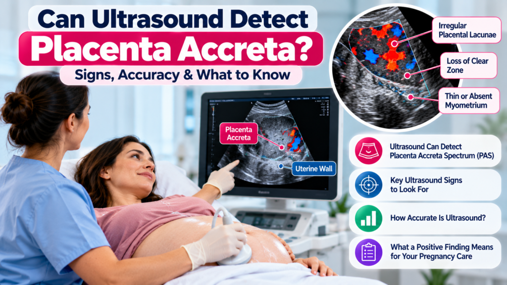
Several ultrasound findings have been associated with placenta accreta spectrum.
1. Placental Lacunae
Placental lacunae are irregular vascular spaces within the placenta.
Multiple, large or irregular lacunae can be an important ultrasound feature of PAS.
On color Doppler, these spaces may demonstrate turbulent or high-velocity blood flow. ACOG identifies multiple lacunae and turbulent flow among the findings strongly associated with PAS.
However, lacunae alone do not prove placenta accreta because placental vascular spaces can also occur in other pregnancies.
2. Loss of the Retroplacental Clear Zone
Normally, a hypoechoic or relatively clear area can be seen between the placenta and myometrium.
Loss or irregularity of this retroplacental clear zone may raise suspicion for PAS.
A 2024 systematic review and meta-analysis found loss of the clear zone to be one of the important ultrasound markers in the second and third trimesters.
3. Very Thin Myometrium
The myometrium behind the placenta may appear extremely thin or disrupted.
ACOG specifically describes retroplacental myometrial thickness of less than 1 mm as an ultrasound abnormality associated with PAS.
4. Abnormal Uterine-Bladder Interface
An irregular or disrupted interface between the uterus and bladder can raise concern for abnormal placental invasion.
This finding is particularly important when there is an anterior low-lying placenta or placenta previa over a previous cesarean scar.
5. Placental or Uterine Bulging
The uterus or placenta may appear to bulge toward the bladder or beyond the normal uterine contour.
This can be particularly concerning when combined with other ultrasound findings.
6. Bridging Vessels
Abnormal vessels may appear to extend from the placenta across the uterine wall toward the uterine serosa or bladder.
These are often evaluated using color Doppler.
Color Doppler Findings in Placenta Accreta
Color Doppler can provide additional information when PAS is suspected.
Possible findings include:
- Turbulent flow within placental lacunae
- Increased subplacental vascularity
- Abnormal vessels crossing the uterine wall
- Bridging vessels
- Increased vascularity around the uterine-bladder interface
- Gaps or abnormalities in normal myometrial blood flow
ACOG notes that color Doppler may facilitate PAS diagnosis, with turbulent lacunar blood flow among the commonly described findings.
Importantly, Doppler findings should be interpreted together with grayscale findings and the patient’s clinical risk factors rather than used as a standalone diagnosis.
Who Is at Higher Risk for Placenta Accreta?
Certain factors substantially increase the risk of PAS.
The most important include:
Previous Cesarean Delivery
A previous C-section is one of the most important risk factors, and risk generally increases as the number of previous cesarean deliveries increases.
Placenta Previa
Placenta previa is particularly important when combined with a previous cesarean delivery.
The combination of an anterior placenta previa or low-lying placenta over a cesarean scar should raise clinical suspicion for PAS.
Other Uterine Surgery
Other procedures that can disrupt the uterine lining or myometrial interface may also increase risk.
Examples include certain uterine surgeries and previous curettage procedures.
Other Risk Factors
Additional factors may include:
- Multiple previous pregnancies
- Advanced maternal age
- Previous uterine procedures
- Prior uterine scarring
- Asherman syndrome
Clinical history should always be considered together with ultrasound findings.
How Accurate Is Ultrasound for Placenta Accreta?
Ultrasound can be highly useful, particularly in pregnancies already considered high risk.
A 2024 systematic review and meta-analysis of 37 studies involving 5,764 pregnancies at risk for PAS reported approximately 88% sensitivity and 92% specificity for ultrasound performed during the second and third trimesters.
Another meta-analysis reported overall ultrasound sensitivity of approximately 87% and specificity of approximately 86% across included studies.
These numbers should not be interpreted as a guarantee for an individual patient. Accuracy can vary depending on:
- Placental location
- Degree of placental invasion
- Previous C-section history
- Gestational age
- Ultrasound equipment
- Sonographer experience
- Patient characteristics
- Whether the pregnancy was already considered high risk
Can a Normal Ultrasound Rule Out Placenta Accreta?
No.
This is one of the most important points for patients and healthcare professionals to understand.
ACOG states that the absence of ultrasound findings does not exclude placenta accreta spectrum. Clinical risk factors remain important even when ultrasound does not show obvious PAS abnormalities.
For example, a patient with placenta previa over a previous cesarean scar may remain at significant risk even if the ultrasound findings are not definitive.
This is why suspected PAS is best assessed using the combination of:
Clinical risk factors + placental location + grayscale ultrasound + color Doppler + expert interpretation.
Can Placenta Accreta Be Seen in the First Trimester?
Yes.
Some PAS-related ultrasound findings can appear during the first trimester, although many cases are recognized later in pregnancy.
A 2024 meta-analysis found that first-trimester ultrasound can detect PAS in pregnancies at risk, with reported sensitivity around 86% and specificity around 63% in the included studies.
One particularly important first-trimester condition is cesarean scar pregnancy, in which the gestational sac implants within or at the site of a previous cesarean scar.
Early recognition is important because cesarean scar pregnancy is strongly associated with subsequent PAS if the pregnancy continues.
When Is MRI Used for Placenta Accreta?
MRI can provide additional information in selected cases, but it is generally not the first-line imaging test for suspected PAS.
MRI may be considered when:
- Ultrasound findings are inconclusive
- The placenta is posterior
- There is concern about the depth or extent of invasion
- Bladder or other organ involvement needs further assessment
- The ultrasound examination is technically limited
Research comparing ultrasound and MRI has found broadly similar diagnostic accuracy, although MRI may provide additional anatomical information in selected difficult cases.
Therefore, MRI is usually considered an adjunct rather than a replacement for expert ultrasound assessment.
What Happens If Ultrasound Suggests Placenta Accreta?
If ultrasound findings and clinical risk factors suggest PAS, the patient may be referred to a maternal-fetal medicine (MFM) specialist or a center experienced in managing placenta accreta spectrum.
The purpose of antenatal diagnosis is not simply to put a label on the ultrasound report.
It allows the medical team to plan:
- Delivery location
- Timing of delivery
- Surgical team
- Anesthesia support
- Blood products
- Possible urology involvement
- Maternal-fetal medicine consultation
- Neonatal care
- Emergency planning
ACOG recommends that suspected PAS be managed in appropriately experienced centers because severe hemorrhage and complex surgery may occur.
Medical Disclaimer
This article is for educational and informational purposes only and is not a substitute for professional medical advice, diagnosis, or treatment. Ultrasound findings should be interpreted by a qualified healthcare professional in the context of the patient’s complete medical history and clinical risk factors.
Sources
- American College of Obstetricians and Gynecologists (ACOG), Placenta Accreta Spectrum
- Society for Maternal-Fetal Medicine (SMFM) guidance on ultrasound assessment for PAS
- Ultrasound in Obstetrics & Gynecology, 2024 systematic review and meta-analysis of trimester-specific ultrasound accuracy
- BMC Pregnancy and Childbirth, systematic review and meta-analysis of ultrasound accuracy
- RadiologyInfo, Obstetric Ultrasound