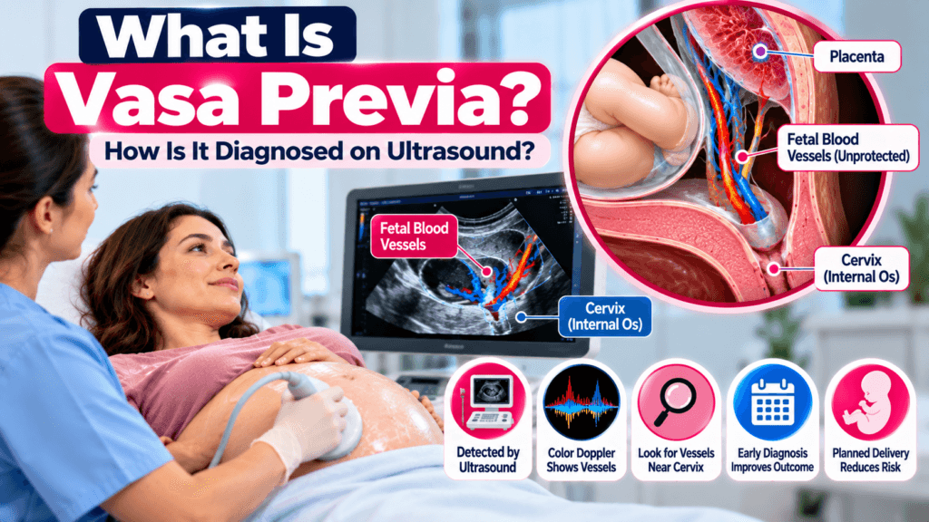
Vasa previa is a rare but potentially serious pregnancy complication in which unprotected fetal blood vessels run through the membranes near or over the internal opening of the cervix. Because these vessels are not protected by the normal cushioning of the umbilical cord or placenta, they can rupture when the membranes break.
The condition is especially important to recognize before labor or rupture of membranes because the vessels contain fetal blood. If they rupture, the baby can lose blood rapidly.
The good news is that vasa previa can often be diagnosed during pregnancy with ultrasound, particularly with transvaginal ultrasound and color Doppler. Prenatal diagnosis allows the obstetric team to plan delivery before labor or membrane rupture, substantially reducing the risk of severe fetal blood loss.
What Is Vasa Previa?
Vasa previa is a condition in which fetal blood vessels travel within the membranes over or near the internal cervical os without the normal protection provided by Wharton’s jelly and placental tissue.
Normally, the umbilical vessels are protected within the umbilical cord.
With vasa previa, vulnerable fetal vessels may travel through the membranes in the lower part of the uterus.
If these vessels are located near the cervix, they may be at risk of being compressed or torn when the membranes rupture.
Because these are fetal vessels, bleeding can represent fetal blood loss rather than maternal blood loss.
Why Is Vasa Previa Dangerous?
The major concern is rupture of fetal blood vessels.
If the membranes rupture and a vessel tears, the fetus can lose blood quickly. Severe fetal hemorrhage can lead to:
- Fetal anemia
- Abnormal fetal heart rate
- Severe fetal blood loss
- Fetal hypoxia
- Emergency delivery
- Stillbirth in severe untreated cases
This is why prenatal diagnosis is so important.
A 2025 systematic review and meta-analysis of standardized screening studies involving more than 779,000 pregnancies found that most confirmed cases of vasa previa were diagnosed before delivery, and reported very high pooled detection performance when standardized second-trimester screening was followed by third-trimester confirmation.
Types of Vasa Previa
SMFM describes two primary types of vasa previa.
Type I Vasa Previa
Type I is commonly associated with velamentous cord insertion.
In velamentous cord insertion, the umbilical vessels travel through the membranes before reaching the placenta instead of remaining protected within placental tissue.
If these exposed vessels travel over or close to the cervix, vasa previa can develop.
Type II Vasa Previa
Type II is generally associated with a succenturiate lobe or bilobed placenta.
In this situation, fetal vessels may travel through the membranes between the main placental portion and an accessory placental lobe.
If these vessels pass near the internal cervical os, they can result in vasa previa.
What Causes Vasa Previa?
Vasa previa is usually related to abnormal placental or umbilical cord anatomy.
Important associations include:
Velamentous Cord Insertion
The umbilical cord inserts into the membranes rather than directly into the placental mass.
The vessels then travel unprotected through the membranes.
Succenturiate Placental Lobe
An additional placental lobe may develop separately from the main placenta.
Connecting fetal vessels can run between the two portions.
Bilobed Placenta
A placenta divided into two relatively distinct lobes can have connecting vessels that travel through the membranes.
Low-Lying Placenta or Placenta Previa
A low-lying placenta or placenta previa, particularly in the second trimester, is associated with increased risk of vasa previa.
SMFM notes that many cases have a history of a low-lying placenta or second-trimester placenta previa.
IVF Pregnancy
Pregnancies conceived through in vitro fertilization have been associated with an increased risk of vasa previa.
Risk Factors for Vasa Previa
Risk factors and associated findings include:
- Velamentous cord insertion
- Succenturiate placental lobe
- Bilobed placenta
- Low-lying placenta
- Placenta previa
- IVF pregnancy
- Multiple pregnancy
- Abnormal placental morphology
- Previous uterine or placental abnormalities
Having a risk factor does not mean that vasa previa is definitely present.
It means the placental and cervical regions may deserve more detailed ultrasound evaluation.
How Is Vasa Previa Diagnosed on Ultrasound?
Ultrasound is the main method used for prenatal diagnosis of vasa previa.
The examination may begin with transabdominal ultrasound.
The sonographer or physician may assess:
- Placental location
- Umbilical cord insertion
- Relationship between the placenta and cervix
- Lower uterine segment
- Internal cervical os
- Fetal vessels near the cervix
When the relationship between vessels and the cervix is uncertain, transvaginal ultrasound with color Doppler provides a more detailed evaluation.
What Does Vasa Previa Look Like on Ultrasound?
The appearance depends on the anatomy, but several findings can raise suspicion.
Fetal Vessel Near the Internal Cervical Os
One of the most important findings is a fetal blood vessel running across or very close to the internal cervical os.
Color Doppler can demonstrate that the structure contains blood flow.
Vessel Traveling Through the Membranes
The vessel may be seen coursing through the membranes rather than being contained within the umbilical cord.
Velamentous Cord Insertion
The umbilical cord may insert into the membranes rather than directly into the placenta.
This finding increases concern for Type I vasa previa if exposed vessels travel toward the cervix.
Vessels Between Placental Lobes
When a succenturiate or bilobed placenta is present, connecting vessels may be seen running between the placental portions.
If these vessels approach the internal os, Type II vasa previa should be considered.
Color Doppler Flow
Color Doppler can confirm that the structure near the cervix is a blood vessel and can help demonstrate its course.
A vessel with fetal blood-flow characteristics crossing or approaching the cervical os is an important diagnostic finding.
Why Is Transvaginal Ultrasound Important?
The internal cervical os and lower uterine segment can be difficult to evaluate completely with transabdominal imaging alone.
Transvaginal ultrasound brings the transducer closer to the cervix and can provide a clearer assessment of vessels near the internal os.
Color Doppler can then be used to determine whether a structure represents a blood vessel and to evaluate its relationship with the cervix.
A systematic review found that ultrasound accuracy for vasa previa diagnosis was particularly high when transvaginal ultrasound was combined with color Doppler. In two prospective studies included in that review, second-trimester transvaginal color Doppler detected all identified cases, although the evidence base was relatively small.
When Is Vasa Previa Usually Diagnosed?
Vasa previa is often identified during the second-trimester anatomy ultrasound, commonly around 18 to 22 weeks.
SMFM notes that diagnosis is often made between approximately 18 and 26 weeks.
A newer international expert consensus published in 2024 recommended that the second-trimester anatomy scan include examination of placental cord insertion and a color Doppler sweep of the region over the cervix. The same consensus recommended transvaginal ultrasound with Doppler around 32 weeks when a low-lying placenta or placenta previa was identified in the second trimester.
However, screening recommendations are not completely uniform across all professional organizations and countries. A 2026 review of international guidelines found considerable variation in whether routine vasa previa screening is explicitly recommended.
Can Vasa Previa Resolve?
Yes, some cases diagnosed in the second trimester may no longer meet criteria later in pregnancy.
SMFM notes that approximately 20% of cases diagnosed in the second trimester may resolve.
This can happen because the placenta and uterus change position and shape as pregnancy progresses.
However, a suspected diagnosis should not simply be assumed to have disappeared.
Follow-up imaging may be recommended to determine whether the fetal vessels remain close to the cervix.
What Happens If Vasa Previa Is Found?
If vasa previa is diagnosed, the pregnancy usually requires specialized obstetric management.
The medical team may recommend:
- Follow-up ultrasound
- Transvaginal color Doppler
- Monitoring for symptoms
- Fetal surveillance
- Planning delivery before labor
- Delivery by scheduled cesarean when appropriate
The goal is to avoid spontaneous labor or rupture of membranes while vulnerable fetal vessels remain near the cervix.
SMFM’s current reaffirmed guidance recommends planned cesarean delivery in the 34 to 37 week range, with the exact timing individualized according to the clinical situation.
The 2024 international expert consensus suggested scheduled cesarean delivery between 35 and 37 weeks for asymptomatic patients with vasa previa.
What If the Water Breaks With Vasa Previa?
Rupture of membranes can be an emergency when vasa previa is present because the fetal vessels may tear.
A pregnant person with known vasa previa should follow the emergency instructions provided by their obstetric team.
Possible warning signs include:
- Vaginal bleeding
- Regular contractions
- Leakage of fluid
- Sudden decrease in fetal movement
- Abnormal fetal heart rate symptoms or concerns
Vaginal bleeding after membrane rupture requires urgent medical evaluation, particularly when vasa previa is known or suspected.
Vasa Previa vs Placenta Previa
These conditions sound similar but are different.
| Condition | What Is Abnormal? |
|---|---|
| Vasa previa | Fetal blood vessels run through the membranes near or over the cervix |
| Placenta previa | Placental tissue covers or lies over the internal cervical os |
| Low-lying placenta | Placenta is close to the cervix but does not necessarily cover the os |
A person can have placenta previa without vasa previa, and vasa previa can occur without placenta previa.
However, placenta previa or a low-lying placenta can increase the risk of vasa previa.
Can ultrasound detect vasa previa?
Yes. Ultrasound can detect vasa previa, particularly when the placental cord insertion and lower uterine segment are carefully evaluated with color Doppler. Transvaginal ultrasound is especially useful when vessels are suspected near the internal cervical os.
What is the best ultrasound for vasa previa?
Transvaginal ultrasound with color Doppler is particularly useful for evaluating fetal vessels near the cervix.
Can vasa previa be seen at the 20-week ultrasound?
Yes. Many cases are diagnosed during the second-trimester anatomy scan, although follow-up may be needed later in pregnancy.
Can vasa previa go away?
Some cases identified during the second trimester may no longer be present later in pregnancy. Follow-up ultrasound is important to determine whether the vessels remain near the cervix.
Does vasa previa always require a C-section?
When vasa previa persists near delivery, planned cesarean delivery is generally recommended to avoid labor and rupture of membranes. The exact timing should be determined by the maternal-fetal medicine or obstetric team.
What causes vasa previa?
Common associations include velamentous cord insertion, succenturiate or bilobed placenta, low-lying placenta, placenta previa, and IVF pregnancy.
Is vasa previa the same as placenta previa?
No. Placenta previa involves placental tissue near or covering the cervix, while vasa previa involves fetal blood vessels traveling near or over the cervix.
Medical Disclaimer
This article is for educational and informational purposes only and is not a substitute for professional medical advice, diagnosis, or treatment. Ultrasound findings should always be interpreted by a qualified obstetrician, maternal-fetal medicine specialist, or appropriately trained ultrasound professional in the context of the complete pregnancy history.
Sources
- Society for Maternal-Fetal Medicine (SMFM), Consult Series #37: Diagnosis and Management of Vasa Previa, reaffirmed 2024.
- 2024 International Expert Consensus on Diagnosis and Management of Vasa Previa.
- 2025 Systematic Review and Meta-analysis of Ultrasound Screening for Vasa Previa.
- Systematic Review of Accuracy of Ultrasound in Diagnosis of Vasa Previa.